|Articles|March 1, 2021
Study Results Show Half of Severe COVID-19 Patients Have Damage to the Heart
Author(s)Gabrielle Ientile
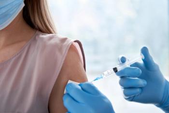
Investigators used MRI scans of the heart to identify whether heart injury was caused by COVID-19 infection.
Advertisement
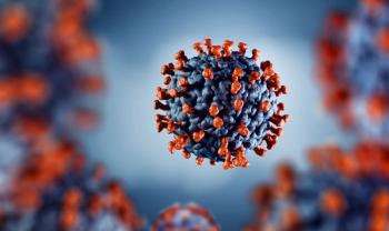
Approximately 50% of patients hospitalized for severe COVID-19 and with elevated troponin levels presented lasting heart damage, including myocarditis, infarction, ischemia, as well as combinations of all 3, according to a recent study.1,2
Investigators from London, United Kingdom, utilized multi-parametric cardiovascular magnetic resonance (CMR) to diagnose causes of elevated troponin in patients who recovered from SARS-CoV-2.1 The study, published in the European Heart Journal, is the largest of its kind to date.
Acute respiratory infections and sepsis are often indicative of elevated serum troponin levels in hospitalized patients, which may lead to death even after a patient’s recovery, investigators explained.
The study recruited 148 patients with severe infection of the virus, defined as requiring hospitalization, and 48% of which additionally required ventilation support, who experienced troponin elevation up to June 2020 among 6 hospitals across 3 NHS Foundation trusts: Royal Free London, Imperial College Healthcare, and University College London Hospital.
COVID-19 infection was diagnosed through either:
- a positive combined oro/nasopharyngeal throat swab or tracheal aspirate for SARS-CoV-2 by reverse-transcriptase-polymerase chain reaction (PCR), or
- a negative SARS-CoV-2 swab with a demonstration of illness, including cough, fever, myalgia, as well as typical blood biomarkers and high likelihood of viral infection.
Following hospital discharge, the 148 patients with an abnormal high-sensitivity troponin (hsTnT14 ng/L for Royal Free and UCLH; hsTnI14 ng/L for females and 30 mL/min/m2 for males for Imperial) underwent CMR scans. All volunteer participants did not have symptoms or a history of cardiovascular disease or hypertension, and the control group was matched for age, gender, diabetes, and hypertension but with no clinical suspicion of myocardial injury, including myocardial infarction (MI) or myocarditis.
Of the 148 patients, 133 underwent a full tissue characterization protocol (LGE, T1 and T2 mapping). Investigators reported that the mean age of patients was 64 12 years, with 70% of patients being male (104). Eighty-six percent (128) of patients were PCR-positive, and 93% (138) presented typical symptoms of COVID-19. Additionally, the median length of stay in the hospital was 9 days; 32% (48) of patients required admission to the intensive care unit in order to receive ventilation support.
As for cardiac function, left ventricular dysfunction was present in 11% (17) of patients, with 9 cases a result of myocardial infarction, 6 of whom had a history of ischemic heart disease. The average right ventricular ejection fraction (RVEF) was 61% 9% and was the same in the healthy volunteers (RVEF 61% 5%, P = 0.85); however, RVEF was lower in the matched control group (RVEF 64% 7%, P=0.032).
After follow up of at least 1 month, scarring or injury to the heart muscle was found in 54% (80) of patients, stemming from inflammation (26%), infarction or ischemia (22%), or both (6%), with 8% of patients showing ongoing heart inflammation, according to investigators.
"These findings give us 2 opportunities: firstly, to find ways of preventing the injury in the first place, and from some of the patterns we have seen, blood clotting may be playing a role, for which we have potential treatments,” said co-lead researcher Marianna Fontana, professor of cardiology at University College London, United Kingdom.2 “Secondly, detecting the consequences of injury during convalescence may identify subjects who would benefit from specific supporting drug treatments to protect heart function over time.”
Study authors stipulated that it remains uncertain whether the findings demonstrate preexisting disease or that brought on by COVID-19, and more research is necessary.
“We found evidence of high rates of heart muscle injury that could be seen on the scans a month or 2 after discharge. Whilst some of this may have been preexisting, MRI scanning shows that some were new, and likely caused by COVID-19,” Fontana said.2
“Importantly, the pattern of damage to the heart was variable, suggesting that the heart is at risk of different types of injury. While we detected only a small amount of ongoing injury, we saw injury to the heart that was present even when the heart's pumping function was not impaired and might not have been picked up by other techniques. In the most severe cases, there are concerns that this injury may increase the risks of heart failure in the future, but more work is needed to investigate this further."
References
1. Kotecha T, Knight DS, Razvi Y, et al. Patterns of myocardial injury in recovered troponin-positive COVID-19 patients assessed by cardiovascular magnetic resonance. European Heart Journal; February 18, 2021. doi:
2. Damage to the heart found in more than half of COVID-19 patients discharged from hospital. News release. European Society of Cardiology; February 18, 2021. Accessed February 24, 2021. https://www.escardio.org/The-ESC/Press-Office/Press-releases/Damage-to-the-heart-found-in-more-than-half-of-COVID-19-patients-discharged-from-hospital
Advertisement
Related Content
Advertisement


Latest CME


Advertisement
Advertisement
Trending on Drug Topics
1
ICER Report Finds GLP-1s Are Cost-Effective But Warns of Budget Strain
2
New Study Finds Compounded GLP-1s Remain Robust Despite End of Shortages
3
Once Weekly Oral HIV Regimen Maintains Suppression Through Week 48
4
Postpartum Mothers Favor Bedtime Stopping Rule Over Stricter Chrononutrition Approaches
5