Cover Story: STICKS AND KIDNEY STONES
As low-carb, high-protein diets grow in popularity, the risk of developing kidney stones rises as well.
As low-carb, high-protein diets grow in popularity, the risk of developing kidney stones rises as well.
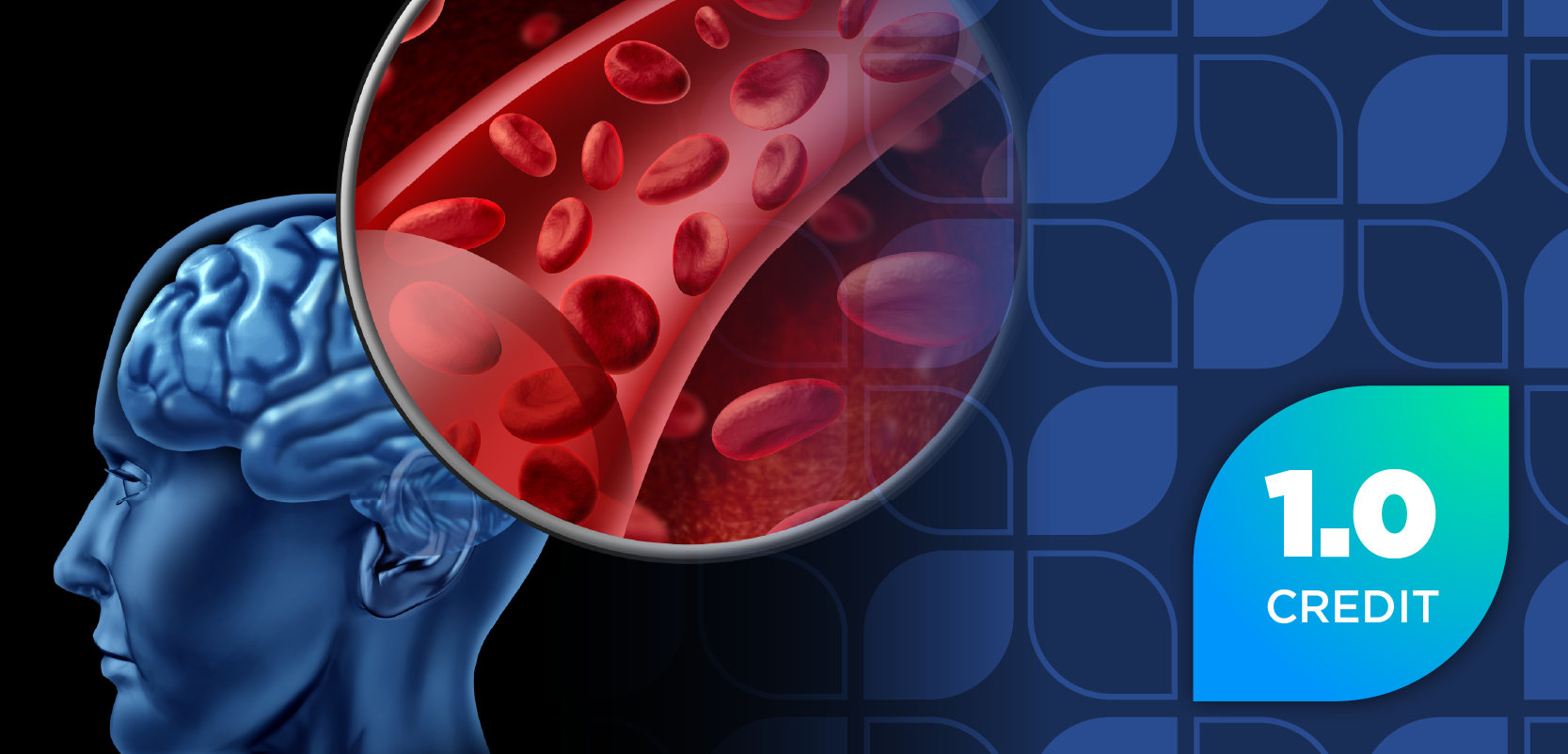
Researchers have learned a great deal about managing kidney stone disease in recent years. But the condition itself has been around for a long time, with evidence of its existence as far back as the ancient Egyptians, whose 7,000-year-old mummies were found with the hardened minerals still lodged within them.
Also called nephrolithiasis, the development of kidney stoneswhich range in size from that of a grain of sand to a golf ballis one of the most common disorders of the urinary tract, affecting about 10% of the U.S. population at some point. Caucasian adults between the ages of 30 and 50 are more likely to develop stones, and men are more prone than women, at a ratio of three to one.
Are diets really to blame?
Recent evidence suggests that the incidence of stone disease in women is increasing at a faster rate than in men, specifically over the past 10 years, said Leslie Spry, M.D., a Nebraska nephrologist and spokesman for the National Kidney Foundation (NKF). "It has been conjectured that this could be the result of recent fad diets, such as high-protein diets."
A study examining the dietary intake of 10,000 nurses over a 10-year period showed that women in the highest quintile of protein intake had significantly more stone disease than those in the lowest quintile, said Spry. This does not imply cause and effect, he admitted, but merely the association. Another study showed high-protein diets caused abnormalities in the urine that would make stone formation more likely, he added.
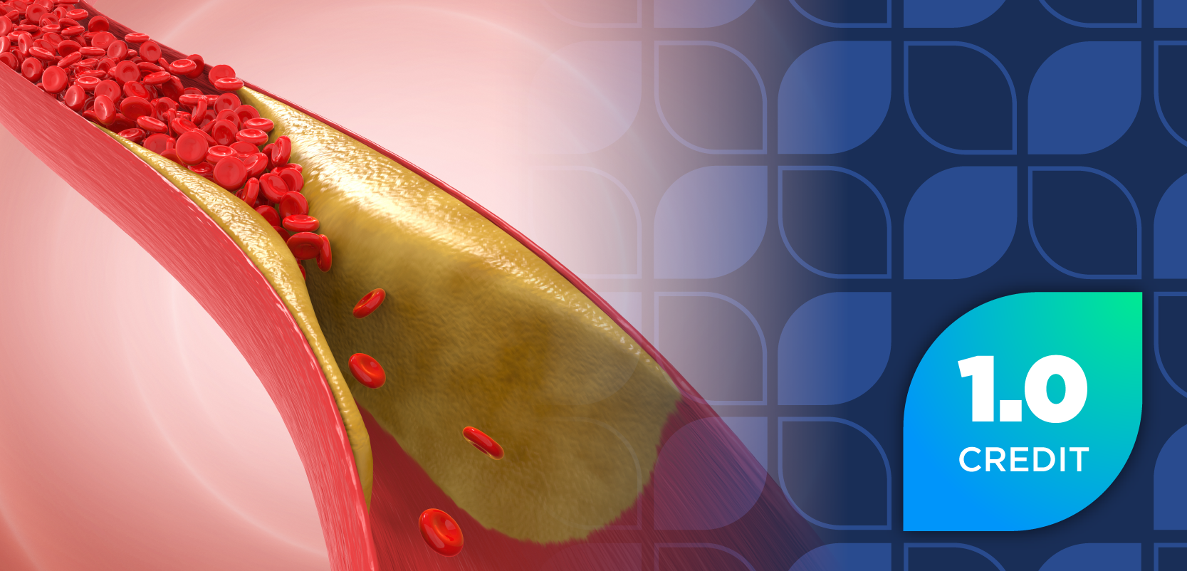
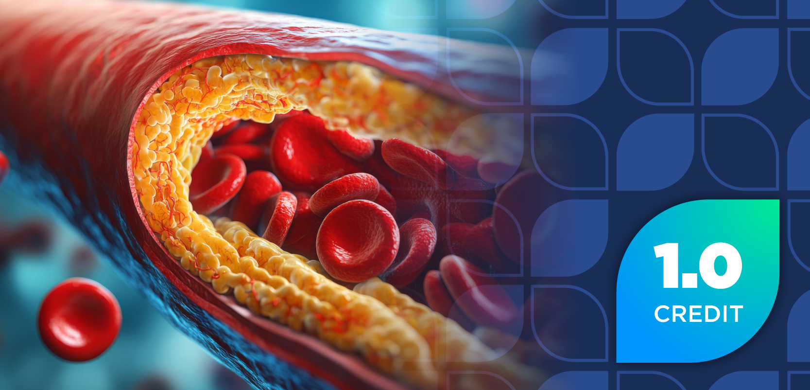
A high-protein load leads to an increase in the glomerular filtration rate of the kidneys, causing the organs to work harder to filter materials at a faster rate. This response can result in elevated blood-urea-nitrogen (BUN) levels and lowered urinary pH from the added uric acid load to the kidneys. The resulting acidic environment promotes calcium excretion. When a diet is also low in carbohydrates, these effects can be enhanced and the combination can lead to the formation of kidney stones, especially in patients with preexisting kidney disease or the elderly, who have a natural decline in kidney function.
According to Gary Curhan, M.D., nephrologist at Brigham and Women's Hospital in Boston and associate professor of medicine at Harvard Medical School, only a diet high in animal protein would increase the risk. Meat protein generates more acid and has a higher sulfur content than vegetable protein. In fact, a study from Italy showed that increasing vegetable protein and decreasing animal protein and salt in the diets of men led to a reduction in the recurrence of kidney stones.
With names such as Atkins, South Beach, Calories Don't Count, Scarsdale, and Stillman, high-protein, low-carb diets are also similar in that they promote urine output by causing a diuretic effect that creates a false sense of weight loss. Typically during the first two weeks, body fat is burned as the body tries to generate energy from noncarbohydrate sources. This leads to the formation of ketone bodies that are released into the bloodstream.
Ketosis, a metabolic state that can be deadly for diabetes patients and dangerous for pregnant women, causes the delivery of many nonreabsorbable molecules to the kidneys, eliciting an osmotic diuretic effect and the mobilization of glucose from glycogenleading to loss of body water. This explains the rapid weight loss that usually occurs at the beginning of dieting and provides another mechanism by which urinary pH is lowered and uric acid levels increased, creating a favorable environment for the formation of stones.
Despite their effect on urine composition, fad diets are only one factor in the development of kidney stones. Experts note that there are multiple types of stones that can occur and many underlying disorders that can put one at risk for forming them. People with gout, inflammatory bowel disease, high blood pressure, urinary tract infections, chronic diarrhea, kidney disease, leukemia, lymphoma, multiple myeloma, sarcoidosis, hyperparathyroidism, lead toxicity, and a family history of kidney stones and those who are obese are at increased risk of developing stones.
"A higher body mass index increases the risk of stones," said Curhan. Obesity is associated with abnormal blood and urine chemistries that can lead to stone development. In fact, in 2003, the American Urological Association (AUA) encouraged physicians to make a strong effort to evaluate their obese patients for stone risk and, when needed, institute appropriate medical and dietary therapy to correct metabolic abnormalities such as gouty diathesis, hypercalciuria, and hyperuricosuria.
Other risk factors include consuming foods high in oxalate (e.g., strawberries, chocolate, nuts, spinach, cola), sodium, or purines (e.g., animal protein); male gender; binge drinking; fasting; stress, possibly due to the release of vasopressin, which concentrates urine; and sleep position, with stone incidence greater on the side of the body favored during sleep.
Stone formation
According to basic nephrology, in order to remove waste products, the kidneys must be adequately hydrated. If dehydration should occur, substances such as calcium, oxalate, and uric acid cannot dissolve completely, and crystals form that can build up into kidney stones. The urine normally contains inhibitors such as citrate, magnesium, pyrophosphate, and nephrocalcin that prevent crystallization. But when levels of these inhibitors are low, stones are more likely to form, especially when urine pH is below 5.5. This process is called supersaturation.
Sometimes a stone forms through nucleation, a process by which sodium hydrogen urate, uric acid, and hydroxyapatite crystals form a nucleus to which calcium and oxalate ions adhere to form a mixed-composition stone.
The American Foundation for Urologic Disease (AFUD) says that the type of stone that forms is determined by the chemical imbalance present in the urine. About 80% of stones are calcium-based, made up of calcium oxalate, calcium phosphate, or a mixture of the two. Calcium oxalate is the most common form. The calcium phosphate type of stone typically occurs in patients with metabolic or hormonal disorders, including hyperparathyroidism or renal tubular acidosis, in which the kidney is unable to excrete acid and citrate levels get very low. Increased intestinal absorption of calcium (absorptive hypercalcemia) and a kidney defect that allows excessive calcium to enter the urine (renal calcium leak) can also lead to hypercalciuria.
Another 10% of renal stones are made up of uric acid, the end product of purine metabolism, and are more common in men. Whether or not a uric acid stone will form depends upon the pH of the urine. In pH less than 5.5, uric acid crystals will precipitate, and in higher pH (alkaline), the uric acid will remain soluble.
Cystine, an amino acid and a component in 1% of stones, does not dissolve well in urine, according to the National Institute of Diabetes & Digestive & Kidney Diseases (NIDDK). Cystinuria is a rare, autosomal recessive disorder that causes increased urinary excretion of cystine and the formation of difficult-to-treat stones.
A small percentage of stones are known as "infection stones," or struvite stones; they are made of magnesium ammonium phosphate. More common in women, these stones develop after a urinary tract infection has altered the chemical balance of the urine, the pH is above 7.2, and ammonia is present in the urine. Caused by bacteria that produce urease, the enzyme breaks down the urea to form ammonia, bicarbonate, and carbonate ion. Struvite stones can develop jagged edges, known as "staghorns," and can become quite large.
Stone-provoking medications that can cause or exaggerate nephrolithiasis include acetazolamide, calcium-channel blockers, vitamin C, triamterene, calcium with vitamin D, uricosuric agents, calcitriol, probenecid, aspirin, furosemide, theophylline, thyroid hormones, trimethoprim/sulfamethoxazole, acyclovir, indinavir (Crixivan, Merck), topiramate (Topamax, Ortho-McNeil), and other drugs that change the acidity of the urine for long periods of time.
Painful presentation
"The worst pain of my life," is the way Curhan's patients describe how they feel when they arrive in his office. It's typically a deep, unrelenting pain in the side that fluctuates in intensity but never completely goes away until the stone passes, he explained. The pain, also called renal colic, often starts suddenly when the stone moves into the urinary tract. Patients may feel a sharp, cramping pain in the back, side, or lower abdomen that later moves to the groin. However, the severity of pain does not relate directly to the size of stone, according to AFUD. Urologists agree that a very tiny stone with sharp edges can cause more discomfort than a larger, rounded stone.
Patients often cannot find a comfortable position and may pace or move from a standing to sitting or reclining position in search of relief. Nausea and vomiting, blood in the urine, and cloudy or foul-smelling urine are common symptoms, according to NKF. Patients seeking medical help may be sweating and have rapid heart and respiration rates when they arrive. The severe discomfort can also cause a rise in blood pressure. Fever and chills may indicate an infection and can be serious when combined with kidney stone obstruction, requiring treatment with antibiotics. As the stone passes down the ureter and reaches the urinary bladder, patients may experience urgency, frequency, and burning upon urination. Sometimes a stone in this position is mistaken for a urinary tract infection.
In addition to taking a medical history and conducting a physical examination, urologists may perform imaging techniques to establish the presence of a kidney stone and to determine whether it's obstructing the urinary tract. Equally important is determining the substance responsible for forming the stone so that appropriate treatment and preventive measures can be taken. According to NIDDK, this can be accomplished through laboratory blood chemistries, urinalysis, and by examining the stone, if possible. Patients with their first stone should be assessed to find the cause and to identify those at high risk for recurrence.
A rock and a hard place
Treatment of kidney stones involves two distinct phases: the acute phase and the preventive long-term phase that comes after the acute phase has resolved. The cornerstone of therapy for renal colic, once diagnosed, is analgesia (see Table 1). Morphine sulfate is the narcotic drug of choice for parenteral use. Ketorolac tromethamine is sometimes used as an alternative, or in combination with the morphine, to provide pain relief. Oral combinations of narcotics and acetaminophen (APAP), including oxycodone 5 mg/APAP 325 mg and hydrocodone 5 mg/APAP 500 mg, are typically used in the outpatient setting.
Table 1
Drugs to treat renal colic
The calcium-channel blocker nifedipine can relax ureteral smooth muscle and help to facilitate stone passage. Prednisone is added to reduce ureteral inflammation. Use of both nifedipine and prednisone should be limited to five to 10 days. Metoclopramide or prochlorperazine may be given to control nausea and vomiting.
Stone free
Once the stones have been removed through lithotripsy or surgery or passed through the urine, prevention of recurrence becomes the focus of therapy. Because the recurrence rate is 50% within five to 10 years of the first attack, this phase of treatment is very important, according to NIDDK.
Following the first episode of kidney stones, preventive treatment may be conservative and include only diet modification and increased fluid intake. But it is always important to correct any underlying disorder that may be causing stones to form in the first place, recommends AFUD. It is also advisable to treat patients who initially form stones in their teens and twenties aggressively because they're more likely to experience recurrence.
Along with restricting dietary intake of sodium and oxalate, stone-formers should be encouraged to drink plenty of water, enough to cause them to get up and pass urine at least once in the night, recommended NKF's Spry.
Other therapies that are sometimes helpful include:
Diuretics: Used to reduce urinary calcium excretion by increasing proximal and distal tubular absorption of calcium, thiazide diuretics, such as hydrochlorothiazide (HCTZ), chlorothiazide, trichlormethiazide, and chlorthalidone, have been shown to protect the kidneys from stones. "Many patients with elevated urine calcium need HCTZ at doses higher than those needed to treat hypertension," Curhan said. "The typical patient will require 50 mg of HCTZ per day, and many will require as much as 50 mg twice daily."
Thiazides must be used with caution, however, because they also cause loss of potassium, which can, in turn, reduce citrate levels and increase the risk for stones. When thiazides are not effective, potassium-sparing amiloride can be used.
Citrate salts: Citrate salts are used for the prevention of calcium oxalate and uric acid stones because of their ability to inhibit stone formation. Citrate decreases supersaturation of urinary calcium by forming a complex with calcium ions; it can also prevent a stone from getting larger by inhibiting calcium binding on the surface of a crystal that has already formed.
Mission Pharmacal, a manufacturer with several drugs and diagnostic tests on the market for the treatment and prevention of kidney stones, states that its potassium citrate product, Urocit-K, has been proven to inhibit the formation of most stones in more than 90% of patients.
"Urocit-K is particularly effective in patients with renal tubular acidosis since it averts acid excess, raises urinary citrate, and lowers urinary calcium," said Charles Y. C. Pak, M.D., director of the Center for Mineral Metabolism and Clinical Research at the University of Texas Southwestern Medical Center. He developed the drug in his laboratory and later transferred the New Drug Application to Mission.
Patients who develop calcium stones as a result of impaired intestinal absorption due to small bowel disease should be given magnesium citrate. A potassium magnesium citrate combination may be more effective than either potassium or magnesium citrate alone.
Citrate salts should not be used for patients with struvite stones, added Pak. "The bacterial organisms causing the infection can destroy citrate," he explained. Additionally, citrates should not be used for patients with peptic ulcer disease, urinary tract infections, or bleeding disorders. Patients taking potassium citrate should not take any other potassium-sparing or potassium-containing product.
Phosphates: Phosphates are useful in the prevention of calcium stones because they bind calcium and reduce its intestinal absorption, thereby reducing urinary calcium. Potassium phosphate, which can cause diarrhea, is usually taken four times daily after meals to prevent kidney stones. Cellulose phosphate (Calcibind, Mission Pharmacal) should be used only for severe hypercalciuria due to excessive absorption of calcium in the intestine.
Pak, who is also the lead author of Mission Pharmacal's guide for health professionals, entitled ABCs of Medical Management of Stones, warns that cellulose phosphate can bind magnesium (requiring supplementation) and increase oxalate (necessitating rigid dietary oxalate restriction). There is also a concern, he said, that in very high doses, cellulose phosphate may reduce calcium absorption so low that bone loss occurs, and this limits use. "If it is used, bone density should be monitored."
Cholestyramine: By binding oxalate in the intestine, cholestyramine reduces levels in the urine. Deficiencies of fat-soluble vitamins may result with long-term use, and supplements may be necessary.
Pyridoxine: Pyridoxine (vitamin B6) decreases endogenous oxalate production but should not be used in patients with decreased renal function.
Allopurinol: Very effective in reducing the secretion of uric acid required for the formation of a uric acid stone, allopurinol's side effects, such as leukopenia, skin rashes, thrombocytopenia, diarrhea, headache, and fever, may limit its use. Allopurinol will not prevent calcium stones from forming. The rapid reduction in uric acid levels caused by allopurinol may trigger an attack of gout in some people. To offset this possibility, patients should also take a nonsteroidal anti-inflammatory drug for two to three months.
Acidifiers: Renacidin (Guardian Laboratories) and other acidifiers may be used as an irrigation of the urinary tract four to five days after surgery in an attempt to remove apatite and struvite stone fragments. Patients must have healthy kidneys and sterile urine in order to be candidates for irrigation. Acetohydroxamic acid (Lithostat, Mission Pharmacal), which blocks the enzymes released by the bacteria in struvite stones, is sometimes prescribed in combination with antibiotics. Its use is limited, however, due to its side-effect profile. Iron supplements may be necessary due to acetohydroxamic acid's potential to reduce iron stores in the body. Nausea, vomiting, headache, blood clots in the legs, anxiety, rash, and depression are other adverse effects.
Future approaches for treating kidney stones will include the innovative use of existing drugs and some that are not yet approved, predicted Pak. The combined use of potassium citrate and calcium citrate (Citracal, Mission Pharmacal) may be useful in managing patients in whom calcium supplements are deemed necessary for the prevention of bone loss. He added that in Europe they are testing fish oils for the treatment of high urinary calcium and are developing drugs to block oxalate absorption in the bowel.
A summary of medications used to treat and prevent the formation of kidney stones can be found in Table 2.
Table 2
Drugs to treat nephrolithiasis
Can R.Ph.s help?
Pharmacists can play a big role in the management of kidney stones, noted Curhan. For those patients who have had stones, pharmacists can remind them that recurrences can be prevented, he said. For those on medication, the pharmacist can explain that follow-up evaluations with their urologist are necessary to ensure they are receiving the appropriate medication and dose, he added.
"Pharmacists can encourage patients who are taking medication for stone disease to stay on a low-salt and normal-calcium diet," said Spry. Diets low in calcium or dairy products are no longer recommended and, in fact, have been shown to lead to osteoporosis and increased incidence of stone disease (see the "
Heidi Belden, Pharm.D.
For years, it was recommended that patients suffering with calcium-based kidney stones should limit their intake of dietary calcium in an attempt to curb stone formation. More recently, however, researchers have found that cutting back on calcium can actually worsen stone disease because more oxalate is absorbed in the gastrointestinal tract when there is insufficient calcium to bind with it.
A large, prospective study published in the New England Journal of Medicine in 1993 found that high calcium intake decreases the risk of symptomatic kidney stones. The study also determined that patients consuming less than 850 mg of calcium per day had a higher incidence of stones. Additionally, limiting calcium in patients with calcium stones associated with resorption, in which bone is broken down and calcium is released into the blood, can actually promote further bone loss and development of osteoporosis.
"A disproportionate number of stone-formers have decreased bone density," said Gary Curhan, M.D., nephrologist at Brigham and Women's Hospital in Boston. About 50% of women who form calcium-based stones have high urinary calcium excretion. If their dietary calcium is inadequate, they'll be in negative calcium balance, increasing the risk for osteoporosis. Curhan also believes that diets that make an individual prone to stone formation are also associated with adverse effects on bone. High-protein diets may play a role by elevating blood-urea-nitrogen (BUN) levels and lowering urinary pH, creating an acidic environment that promotes calcium excretion and bone resorption.
Jaeger et al. (1994) compared patients who had calcium-containing kidney stones with normal subjects for incidence of bone fractures and found that the stone-formers had lower bone density and more fractures than the normal subjects. Interestingly, this same group of stone-formers consumed a diet with less calcium and more salt and animal protein than the group with better bone quality and fewer fractures.
In general, experts recommend that patients avoid extremes of calcium intake. Dietary calcium should not be restricted if bone disease is present or suspected. The exception is patients with absorptive hypercalciuria type II (AH-II), who may respond to dietary calcium restrictions. Supplementation with calcium is controversial, although one study indicates daily doses as high as 1,200 mg are safe. Doses of calcium above 2,000 mg per day should be avoided.
Newsletter
Pharmacy practice is always changing. Stay ahead of the curve with the Drug Topics newsletter and get the latest drug information, industry trends, and patient care tips.